If you've been hearing a lot about the wonders of the pelvic floor and want to take a closer look at this amazing part of your anatomy, you're in the right place. This handy mini-guide will give you a deeper understanding of the key elements of your pelvic floor. Let's get right to it!
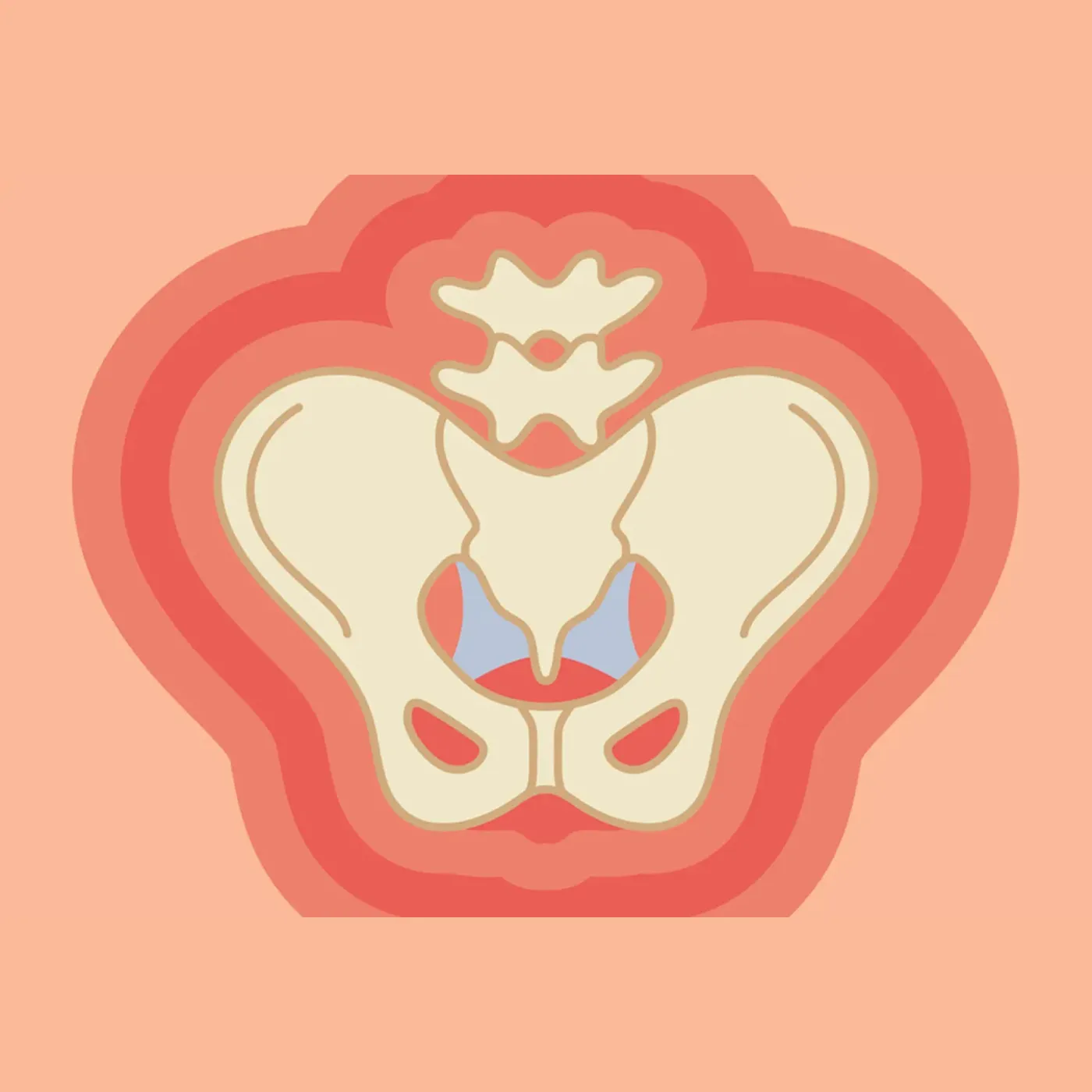
The majority of your pelvic floor is made up of layered muscles that run from your pubic bone to your tailbone, left and right between your two sit bones, and encircle your pelvic openings (vagina, rectum, and urethra). To understand how it all fits together, take a look at the diagram of the pelvic floor below (imagine you're looking down into your pelvic floor from above).

Pelvic Floor Anatomy 101
Now that you have the general lay of the land, let's dive deeper into some key parts of the pelvic floor.
Pubic symphysis
The pubic symphysis is the sturdy joint in the front of your pelvis that's formed by the right and left pubic bones. It’s designed for stability, shock absorption, and moves very little — except in pregnancy, when its width can grow up to 2-3 mm, on average, to prep for birth. Several muscles of the pelvic floor and abdominal muscles attach to areas around this critical joint.
Vagina
This part needs no introduction — this highly flexible, fibromuscular tube runs from the vaginal opening in the vulva to the cervix, passing through an opening of the levator ani muscles along the way.
Sacrum
The triangular bone at the bottom of your spine, connecting your back to your pelvis. The sacrum provides many important attachment sites for your pelvic floor and hip muscle, lending support to your pelvis. The sacrum also provides pathways for several important nerves that control bowel, bladder, and sexual function.
Obturator internus muscle
The most mischievous hip muscle — while most hip muscles are found outside of the pelvis, this one sits largely inside of it. Like other deep hip muscles, the obturator internus supports hip movement, but because it shares connective tissues with the pelvic floor muscles, it can contribute to a host of pelvic floor symptoms.
Rectum
The bottom end of the large intestines collects and holds stool until you are ready to let it out.The rectum passes through the levator ani muscles of the pelvic floor as it forms the anus.
Levator ani muscles
MVP alert! The most important group of muscles of the pelvic floor form a sling of support at the bottom of the pelvis. The levator ani is made up of the puborectalis, pubococcygeus, and iliococcygeus muscles. It runs from the pubic bones to the tailbone and sacrum, and encircles the urethral, vaginal, and anal openings.
Piriformis muscle
The small, deep, hip muscle at the back of your pelvis that helps with the movement and stability of your hips and sacrum. Important nerves from the low back tuck under this muscle before moving on down the legs, making the piriformis a critical muscle to consider when having low back or pelvic pain symptoms.